

Which Dna Strand Is More Often Template
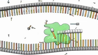
Effigy 2: RNA polymerase (green) synthesizes a strand of RNA that is complementary to the Deoxyribonucleic acid template strand below information technology.
Once RNA polymerase and its related transcription factors are in place, the single-stranded Dna is exposed and fix for transcription. At this point, RNA polymerase begins moving downward the Dna template strand in the 3' to 5' direction, and every bit it does so, it strings together complementary nucleotides. By virtue of complementary base- pairing, this action creates a new strand of mRNA that is organized in the 5' to iii' direction. As the RNA polymerase continues down the strand of Dna, more nucleotides are added to the mRNA, thereby forming a progressively longer concatenation of nucleotides (Figure ii). This process is called elongation.

Figure 3: DNA (top) includes thymine (red); in RNA (lesser), thymine is replaced with uracil (xanthous).
Three of the 4 nitrogenous bases that make upwards RNA — adenine (A), cytosine (C), and guanine (G) — are also found in Dna. In RNA, however, a base called uracil (U) replaces thymine (T) as the complementary nucleotide to adenine (Figure 3). This means that during elongation, the presence of adenine in the DNA template strand tells RNA polymerase to adhere a uracil in the corresponding area of the growing RNA strand (Figure 4).
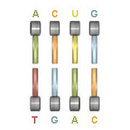
Figure four: A sample section of RNA bases (upper row) paired with Deoxyribonucleic acid bases (lower row). When this base-pairing happens, RNA uses uracil (yellow) instead of thymine to pair with adenine (light-green) in the Dna template below.
Interestingly, this base of operations substitution is not the only divergence between DNA and RNA. A second major deviation between the two substances is that RNA is made in a single-stranded, nonhelical form. (Remember, DNA is almost always in a double-stranded helical grade.) Furthermore, RNA contains ribose sugar molecules, which are slightly different than the deoxyribosemolecules found in Dna. As its name suggests, ribose has more than oxygen atoms than deoxyribose.
Thus, the elongation period of transcription creates a new mRNA molecule from a unmarried template strand of DNA. As the mRNA elongates, it peels abroad from the template as it grows (Effigy 5). This mRNA molecule carries DNA's bulletin from the nucleus to ribosomes in the cytoplasm, where proteins are assembled. However, before information technology can do this, the mRNA strand must divide itself from the Deoxyribonucleic acid template and, in some cases, it must also undergo an editing procedure of sort.
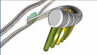
Figure five: During elongation, the new RNA strand becomes longer and longer equally the DNA template is transcribed. In this view, the 5' terminate of the RNA strand is in the foreground. Annotation the inclusion of uracil (yellow) in RNA.

Which Dna Strand Is More Often Template,
Source: http://www.nature.com/scitable/topicpage/the-information-in-dna-is-decoded-by-6524808
Posted by: myersounfee.blogspot.com
0 Response to "Which Dna Strand Is More Often Template"
Post a Comment